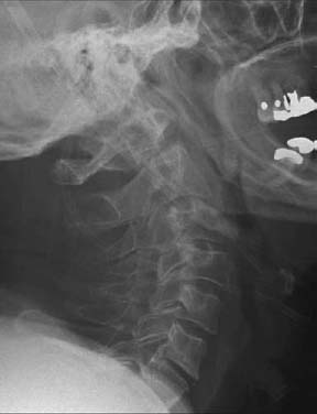
Selection and strict knowledge of the technique. However, surgical success depends on proper patient The literature presents high fusion rates with ORIF (≥80%), which wasĪlso observed in the present study. Concomitant lesions, especially in younger patients, are not One patient presented screw repositioning and one case, dysphonia.ĬONCLUSION: Delayed diagnosis is still an issue in the treatment of odontoid fractures, especially Pseudarthrosis complications were observed in two patients (77% union rate),
#Odontoid fracture complications full#
Patients regained full range of cervical movement, and bone union required approximatelyġ4 weeks. A total of 77% of patients presented a mild or moderate disability (NDI). The pre-operative Frankel score was E in all except one patient (B), inĪ post-operative improvement from B to D was observed. All patients presented type IIb (GrauerĬlassification) fractures, with a mean deviation of 2.95 mm. RESULTS: The mean age of patients was 70 years. Radiologically evaluated (X-ray and CT) pre- and postoperatively. The post-operative cervical range of motion were calculated. Neurological status (Frankel scale) were assessed. METHODS: This was a retrospective study with nine patients. Patients diagnosed with odontoid fracture who underwent open reduction and internal

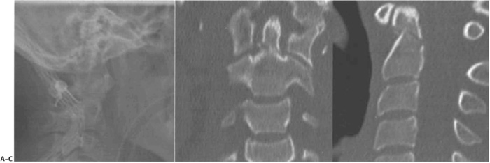
The dens is displaced slightly posteriorly on the body of C2.OBJECTIVE: To evaluate the clinical and radiological outcomes of the surgical treatment in The red arrow points to the same fracture of the sagittal reformatted image. The green arrows point to a transverseįracture of the base of the dens (odontoid) (Type II). These are two reformatted CT images of the cervical spine.


not older than 65 or had a dangerous mechanism of injury, paresthesias in extremities) no fracture of long bone or large laceration) The Canadian criteria (table below) have been reported to be 99.4% sensitive in excluding significant cervical spine pathology There have been studies to examine the need for c-spine imaging in low risk patients.Feeling of instability of head on spine.Inability to move ranging to quadriplegia.Other fractures include a rare longitudinal fracture through dens and body of C2.Unstable fracture as the atlas and occiput can now move together as a unit.Fracture at base of dens at its attachment to body of C2.Classified by location (Anderson and D’Alonzo classification).About 15% of all cervical spine fractures.Dens represents the fused remnants of the body of C1.Transverse ligament holds dens in close approximation to anterior arch of C1.Ligaments (transverse and dentate) provide rotational and translational stability.Odontoid process (dens) of C2 (axis) articulates with C1 (atlas) with three joints.Most odontoid fractures occur with flexion, extension and rotation.As expected, most fatal cervical spine injuries occur at C1 or C2.About 1/3 of C-spine injuries occur at C2 and about ½ at C6-C7.Most dens fractures are caused by motor vehicle accidents and falls.


 0 kommentar(er)
